Draw a Circle Around Four Ball and Socket Joints
Affiliate xix. The Musculoskeletal System
19.three Joints and Skeletal Movement
Learning Objectives
By the end of this section, you will be able to:
- Classify the dissimilar types of joints on the basis of construction
- Explain the role of joints in skeletal movement
The point at which ii or more basic see is called a articulation, or articulation. Joints are responsible for movement, such as the movement of limbs, and stability, such every bit the stability found in the bones of the skull.
Nomenclature of Joints on the Basis of Structure
here are 2 ways to classify joints: on the basis of their structure or on the basis of their office. The structural classification divides joints into bony, fibrous, cartilaginous, and synovial joints depending on the material composing the joint and the presence or absence of a cavity in the joint.
Gristly Joints
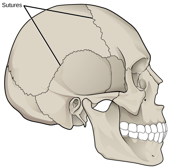
The bones of gristly joints are held together by fibrous connective tissue. There is no cavity, or infinite, present between the basic and and then near gristly joints do not motility at all, or are merely capable of minor movements. There are three types of fibrous joints: sutures, syndesmoses, and gomphoses. Sutures are found merely in the skull and possess brusk fibers of connective tissue that hold the skull bones tightly in place (Figure nineteen.23).
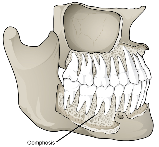
Syndesmoses are joints in which the bones are connected by a ring of connective tissue, allowing for more than movement than in a suture. An instance of a syndesmosis is the joint of the tibia and fibula in the ankle. The amount of movement in these types of joints is determined by the length of the connective tissue fibers. Gomphoses occur between teeth and their sockets; the term refers to the way the tooth fits into the socket like a peg (Figure 19.24). The tooth is connected to the socket by a connective tissue referred to as the periodontal ligament.
Cartilaginous Joints
Cartilaginous joints are joints in which the bones are continued past cartilage. In that location are two types of cartilaginous joints: synchondroses and symphyses. In a synchondrosis, the bones are joined by hyaline cartilage. Synchondroses are institute in the epiphyseal plates of growing basic in children. In symphyses, hyaline cartilage covers the terminate of the bone but the connection between bones occurs through fibrocartilage. Symphyses are constitute at the joints between vertebrae. Either blazon of cartilaginous joint allows for very little motility.
Synovial Joints
Synovial joints are the only joints that take a infinite between the adjoining bones (Figure 19.25). This space is referred to as the synovial (or joint) cavity and is filled with synovial fluid. Synovial fluid lubricates the joint, reducing friction betwixt the bones and allowing for greater movement. The ends of the bones are covered with articular cartilage, a hyaline cartilage, and the entire articulation is surrounded past an articular capsule composed of connective tissue that allows motion of the joint while resisting dislocation. Articular capsules may too possess ligaments that concur the bones together. Synovial joints are capable of the greatest movement of the iii structural joint types; all the same, the more mobile a joint, the weaker the joint. Knees, elbows, and shoulders are examples of synovial joints.
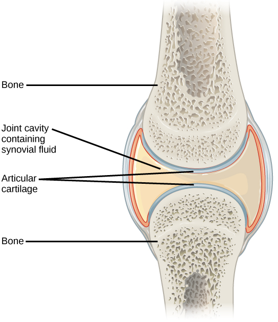
Synovial joints are the only joints that have a infinite or "synovial cavity" in the joint.
Classification of Joints on the Basis of Office
The functional classification divides joints into three categories: synarthroses, amphiarthroses, and diarthroses. A synarthrosis is a articulation that is immovable. This includes sutures, gomphoses, and synchondroses. Amphiarthroses are joints that let slight movement, including syndesmoses and symphyses. Diarthrosesare joints that let for gratis motility of the joint, equally in synovial joints.
Movement at Synovial Joints
The wide range of movement allowed past synovial joints produces dissimilar types of movements. The motion of synovial joints can be classified every bit one of four different types: gliding, athwart, rotational, or special motility.
Gliding Motility
Gliding movementsoccur as relatively flat bone surfaces move past each other. Gliding movements produce very little rotation or angular movement of the bones. The joints of the carpal and tarsal bones are examples of joints that produce gliding movements.
Angular Move
Angular movements are produced when the angle between the bones of a joint changes. There are several different types of angular movements, including flexion, extension, hyperextension, abduction, adduction, and circumduction. Flexion, or angle, occurs when the angle between the basic decreases. Moving the forearm upward at the elbow or moving the wrist to move the mitt toward the forearm are examples of flexion.Extension is the reverse of flexion in that the angle between the bones of a joint increases. Straightening a limb after flexion is an example of extension. Extension past the regular anatomical position is referred to as hyperextension. This includes moving the neck back to look upwards, or bending the wrist then that the manus moves away from the forearm.
Abduction occurs when a bone moves away from the midline of the torso. Examples of abduction are moving the artillery or legs laterally to lift them straight out to the side. Adductionis the movement of a bone toward the midline of the trunk. Motility of the limbs inward later abduction is an example of adduction.Circumduction is the movement of a limb in a round motion, as in moving the arm in a circular motion.
Rotational Movement
Rotational move is the move of a os as it rotates around its longitudinal centrality. Rotation tin can be toward the midline of the trunk, which is referred to as medial rotation, or away from the midline of the body, which is referred to as lateral rotation. Movement of the head from side to side is an example of rotation.
Special Movements
Some movements that cannot be classified equally gliding, angular, or rotational are called special movements. Inversion involves the soles of the feet moving inward, toward the midline of the body. Eversion is the opposite of inversion, movement of the sole of the foot outward, away from the midline of the torso. Protraction is the inductive motion of a bone in the horizontal plane. Retractionoccurs as a articulation moves back into position afterwards protraction. Protraction and retraction tin be seen in the move of the mandible every bit the jaw is thrust outwards and then back inwards. Height is the move of a bone upward, such as when the shoulders are shrugged, lifting the scapulae. Depression is the opposite of elevation—motility downward of a bone, such every bit after the shoulders are shrugged and the scapulae return to their normal position from an elevated position.Dorsiflexion is a angle at the ankle such that the toes are lifted toward the articulatio genus. Plantar flexion is a angle at the ankle when the heel is lifted, such as when standing on the toes. Supination is the movement of the radius and ulna bones of the forearm so that the palm faces forward. Pronation is the contrary motility, in which the palm faces astern. Opposition is the move of the thumb toward the fingers of the same manus, making information technology possible to grasp and hold objects.
Types of Synovial Joints
Synovial joints are further classified into 6 different categories on the basis of the shape and structure of the joint. The shape of the joint affects the type of move permitted by the joint (Figure 19.26). These joints tin be described equally planar, hinge, pivot, condyloid, saddle, or brawl-and-socket joints.
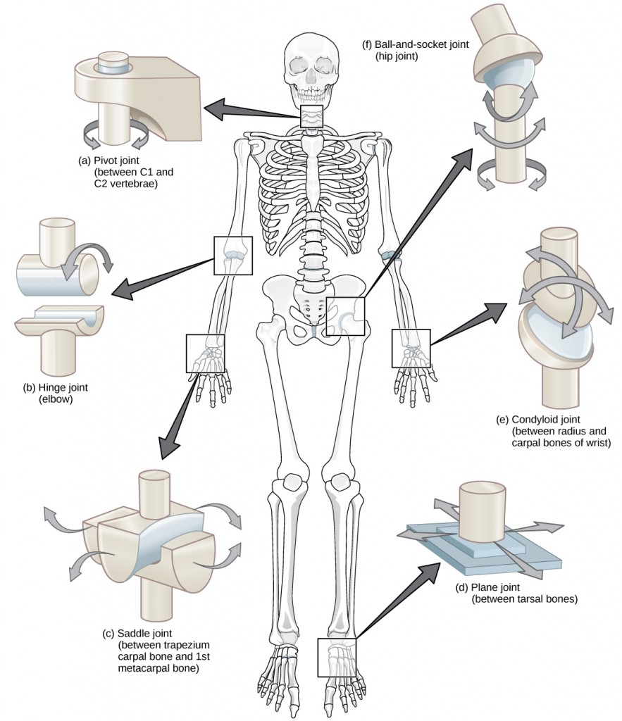
Dissimilar types of joints let different types of move. Planar, hinge, pivot, condyloid, saddle, and ball-and-socket are all types of synovial joints.
Planar Joints
Planar joints have bones with articulating surfaces that are flat or slightly curved faces. These joints let for gliding movements, and so the joints are sometimes referred to every bit gliding joints. The range of motion is express in these joints and does not involve rotation. Planar joints are found in the carpal bones in the hand and the tarsal bones of the human foot, as well as between vertebrae (Effigy 19.27).
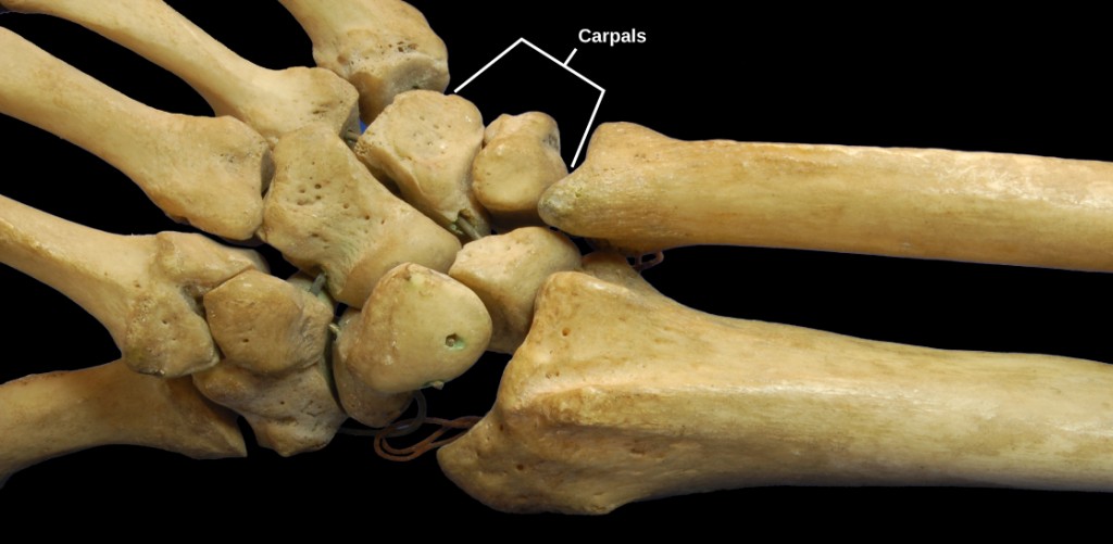
The joints of the carpal bones in the wrist are examples of planar joints. (credit: modification of work past Brian C. Goss)
Hinge Joints
In swivel joints, the slightly rounded finish of i bone fits into the slightly hollow end of the other bone. In this way, one bone moves while the other remains stationary, similar the hinge of a door. The elbow is an example of a hinge joint. The knee is sometimes classified as a modified hinge articulation (Figure 19.28).
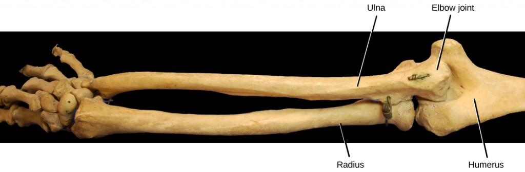
The elbow articulation, where the radius articulates with the humerus, is an case of a hinge joint. (credit: modification of work by Brian C. Goss)
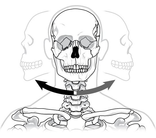
Pivot Joints
Pivot joints consist of the rounded end of one bone fitting into a ring formed by the other bone. This construction allows rotational movement, as the rounded os moves around its own axis. An example of a pivot articulation is the joint of the outset and 2nd vertebrae of the neck that allows the head to move back and forth (Figure 19.29). The articulation of the wrist that allows the palm of the hand to be turned up and down is too a pivot joint.
Condyloid Joints
Condyloid joints consist of an oval-shaped end of ane bone plumbing equipment into a similarly oval-shaped hollow of another bone (Effigy 19.30). This is likewise sometimes chosen an ellipsoidal articulation. This blazon of joint allows angular movement along two axes, every bit seen in the joints of the wrist and fingers, which can move both side to side and up and down.
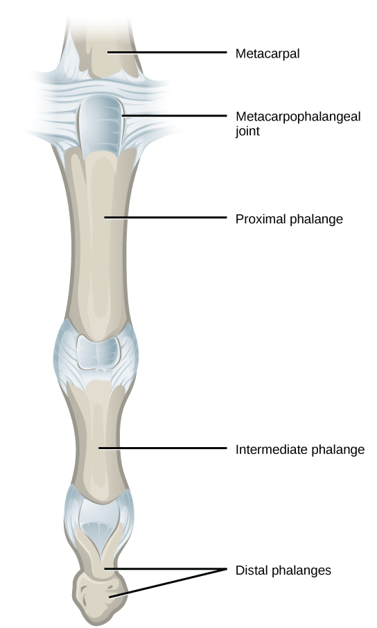
The metacarpophalangeal joints in the finger are examples of condyloid joints. (credit: modification of piece of work by Grey'southward Anatomy)
Saddle Joints
Saddle joints are so named because the ends of each os resemble a saddle, with concave and convex portions that fit together. Saddle joints let angular movements similar to condyloid joints but with a greater range of motion. An instance of a saddle articulation is the thumb joint, which can movement back and forth and up and down, but more freely than the wrist or fingers (Figure 19.31).
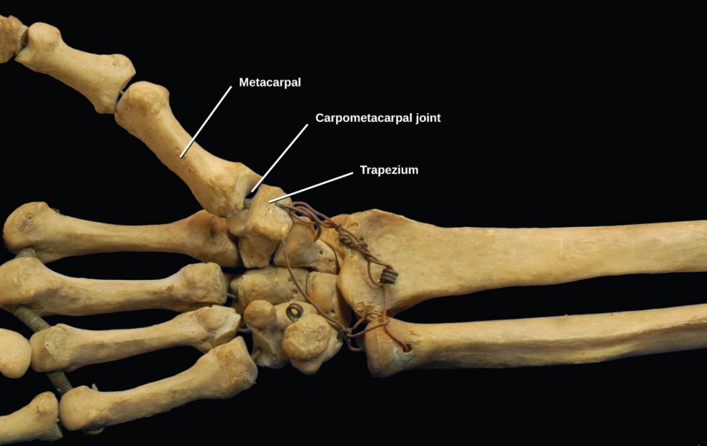
The carpometacarpal joints in the pollex are examples of saddle joints. (credit: modification of work by Brian C. Goss)
Ball-and-Socket Joints
Ball-and-socket joints possess a rounded, ball-like stop of one bone fitting into a cuplike socket of another bone. This system allows the greatest range of motion, as all movement types are possible in all directions. Examples of ball-and-socket joints are the shoulder and hip joints (Figure 19.32).
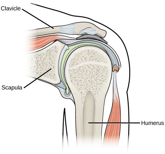
The shoulder joint is an example of a ball-and-socket articulation.
Concept in Activity

Watch this animation showing the 6 types of synovial joints.
Rheumatologist
Rheumatologists are medical doctors who specialize in the diagnosis and treatment of disorders of the joints, muscles, and basic. They diagnose and treat diseases such every bit arthritis, musculoskeletal disorders, osteoporosis, and autoimmune diseases such as ankylosing spondylitis and rheumatoid arthritis.
Rheumatoid arthritis (RA) is an inflammatory disorder that primarily affects the synovial joints of the hands, feet, and cervical spine. Affected joints become swollen, strong, and painful. Although it is known that RA is an autoimmune disease in which the body'due south allowed system mistakenly attacks healthy tissue, the cause of RA remains unknown. Immune cells from the blood enter joints and the synovium causing cartilage breakdown, swelling, and inflammation of the joint lining. Breakdown of cartilage causes bones to rub against each other causing pain. RA is more common in women than men and the age of onset is unremarkably 40–50 years of historic period.
Rheumatologists can diagnose RA on the footing of symptoms such as joint inflammation and hurting, X-ray and MRI imaging, and blood tests. Arthrography is a type of medical imaging of joints that uses a contrast amanuensis, such as a dye, that is opaque to 10-rays. This allows the soft tissue structures of joints—such as cartilage, tendons, and ligaments—to be visualized. An arthrogram differs from a regular 10-ray by showing the surface of soft tissues lining the joint in addition to articulation bones. An arthrogram allows early degenerative changes in joint cartilage to exist detected before basic go afflicted.
There is currently no cure for RA; however, rheumatologists have a number of handling options available. Early stages can exist treated with rest of the affected joints by using a pikestaff or by using joint splints that minimize inflammation. When inflammation has decreased, exercise can be used to strengthen the muscles that surroundings the joint and to maintain joint flexibility. If joint impairment is more extensive, medications can be used to salvage pain and subtract inflammation. Anti-inflammatory drugs such as aspirin, topical pain relievers, and corticosteroid injections may be used. Surgery may be required in cases in which joint damage is astringent.
Summary
The structural classification of joints divides them into bony, fibrous, cartilaginous, and synovial joints. The bones of gristly joints are held together by fibrous connective tissue; the three types of fibrous joints are sutures, syndesomes, and gomphoses. Cartilaginous joints are joints in which the basic are connected by cartilage; the 2 types of cartilaginous joints are synchondroses and symphyses. Synovial joints are joints that take a space betwixt the bordering bones. The functional classification divides joints into iii categories: synarthroses, amphiarthroses, and diarthroses. The motion of synovial joints tin can be classified equally one of four different types: gliding, angular, rotational, or special movement. Gliding movements occur every bit relatively apartment bone surfaces motion by each other. Angular movements are produced when the bending betwixt the bones of a joint changes. Rotational movement is the move of a bone as information technology rotates around its own longitudinal centrality. Special movements include inversion, eversion, protraction, retraction, meridian, depression, dorsiflexion, plantar flexion, supination, pronation, and opposition. Synovial joints are also classified into vi different categories on the footing of the shape and structure of the joint: planar, hinge, pivot, condyloid, saddle, and ball-and-socket.
Exercises
- Synchondroses and symphyses are:
- synovial joints
- cartilaginous joints
- fibrous joints
- condyloid joints
- The motility of bone away from the midline of the body is called ________.
- circumduction
- extension
- adduction
- abductino
- Which of the following is not a characteristic of the synovial fluid?
- lubrication
- daze absorption
- regulation of water residuum in the articulation
- protection of articular cartilage
- The elbow is an example of which type of joint?
- hinge
- pivot
- saddle
- gliding
- What movements occur at the hip articulation and knees as you curve down to bear on your toes?
- What motility(due south) occur(s) at the scapulae when you shrug your shoulders?
Answers
- B
- D
- C
- A
- The hip joint is flexed and the knees are extended.
- Elevation is the movement of a bone upward, such as when the shoulders are shrugged, lifting the scapulae. Depression is the downwards movement of a os, such as later the shoulders are shrugged and the scapulae return to their normal position from an elevated position.
Glossary
- abduction
- when a bone moves away from the midline of the body
- adduction
- motion of the limbs inward after abduction
- amphiarthrosis
- joint that allows slight movement; includes syndesmoses and symphyses
- angular movement
- produced when the angle between the bones of a articulation changes
- articulation
- whatever place where 2 bones are joined
- ball-and-socket joint
- articulation with a rounded, brawl-like end of ane bone plumbing fixtures into a cuplike socket of another bone
- cartilaginous articulation
- articulation in which the bones are connected by cartilage
- circumduction
- movement of a limb in a round motion.
- condyloid joint
- oval-shaped end of ane bone fitting into a similarly oval-shaped hollow of another bone
- depression
- move downwardly of a bone, such every bit subsequently the shoulders are shrugged and the scapulae render to their normal position from an elevated position; contrary of meridian
- diarthrosis
- articulation that allows for gratuitous move of the joint; found in synovial joints
- dorsiflexion
- bending at the ankle such that the toes are lifted toward the knee
- superlative
- motion of a bone upward, such every bit when the shoulders are shrugged, lifting the scapulae
- eversion
- movement of the sole of the foot outward, away from the midline of the body; reverse of inversion
- extension
- motility in which the angle between the bones of a joint increases; opposite of flexion
- fibrous articulation
- joint held together by fibrous connective tissue
- flexion
- move in which the angle betwixt the bones decreases; opposite of extension
- gliding motility
- when relatively flat bone surfaces motility past each other
- gomphosis
- the joint in which the tooth fits into the socket like a peg
- hinge joint
- slightly rounded end of ane bone fits into the slightly hollow terminate of the other bone
- inversion
- soles of the anxiety moving in, toward the midline of the trunk
- articulation
- point at which two or more basic come across
- lateral rotation
- rotation abroad from the midline of the torso
- medial rotation
- rotation toward the midline of the trunk
- opposition
- motion of the pollex toward the fingers of the same mitt, making it possible to grasp and hold objects
- pivot joint
- joint with the rounded terminate of ane bone fitting into a ring formed by the other bone
- planar articulation
- articulation with bones whose articulating surfaces are apartment
- plantar flexion
- angle at the ankle such that the heel is lifted, such equally when standing on the toes
- pronation
- motion in which the palm faces backward
- protraction
- anterior motion of a bone in the horizontal plane
- retraction
- move in which a articulation moves back into position later on protraction
- rotational move
- movement of a bone equally it rotates around its own longitudinal axis
- saddle joint
- articulation with concave and convex portions that fit together; named because the ends of each bone resemble a saddle
- supination
- motion of the radius and ulna bones of the forearm then that the palm faces frontward
- synarthrosis
- articulation that is immovable
- syndesmosis
- joint in which the bones are connected past a band of connective tissue, allowing for more movement than in a suture
- synovial joint
- just joint that has a infinite betwixt the adjoining bones
Source: https://opentextbc.ca/biology/chapter/19-3-joints-and-skeletal-movement/
0 Response to "Draw a Circle Around Four Ball and Socket Joints"
Post a Comment